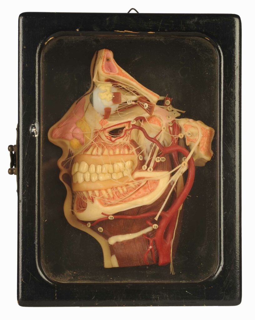
Wax models have been used as training aids for centuries, combining the practices of artistry and medicine to further our understanding of the human body. In this story, Sara studies a German-made wax model of a dissected head made in 1910, and describes how it would have been used to teach dentists and their patients alike.
This model bears the maker’s mark ‘Lehrmittelwerke Berlinische Verlagsanstalt,’ a Berlin-based company that exported their anatomical wax models around the world up to the 1930s at least. The first wax models of the human body were produced during the Renaissance, when they were used by artists seeking to imitate realistic human forms and by medical students, whose education would have otherwise relied upon the availability of fresh corpses for dissection. As medicine became steadily more scientific and the need for intimate anatomical knowledge increased, these models grew in popularity as educational tools.
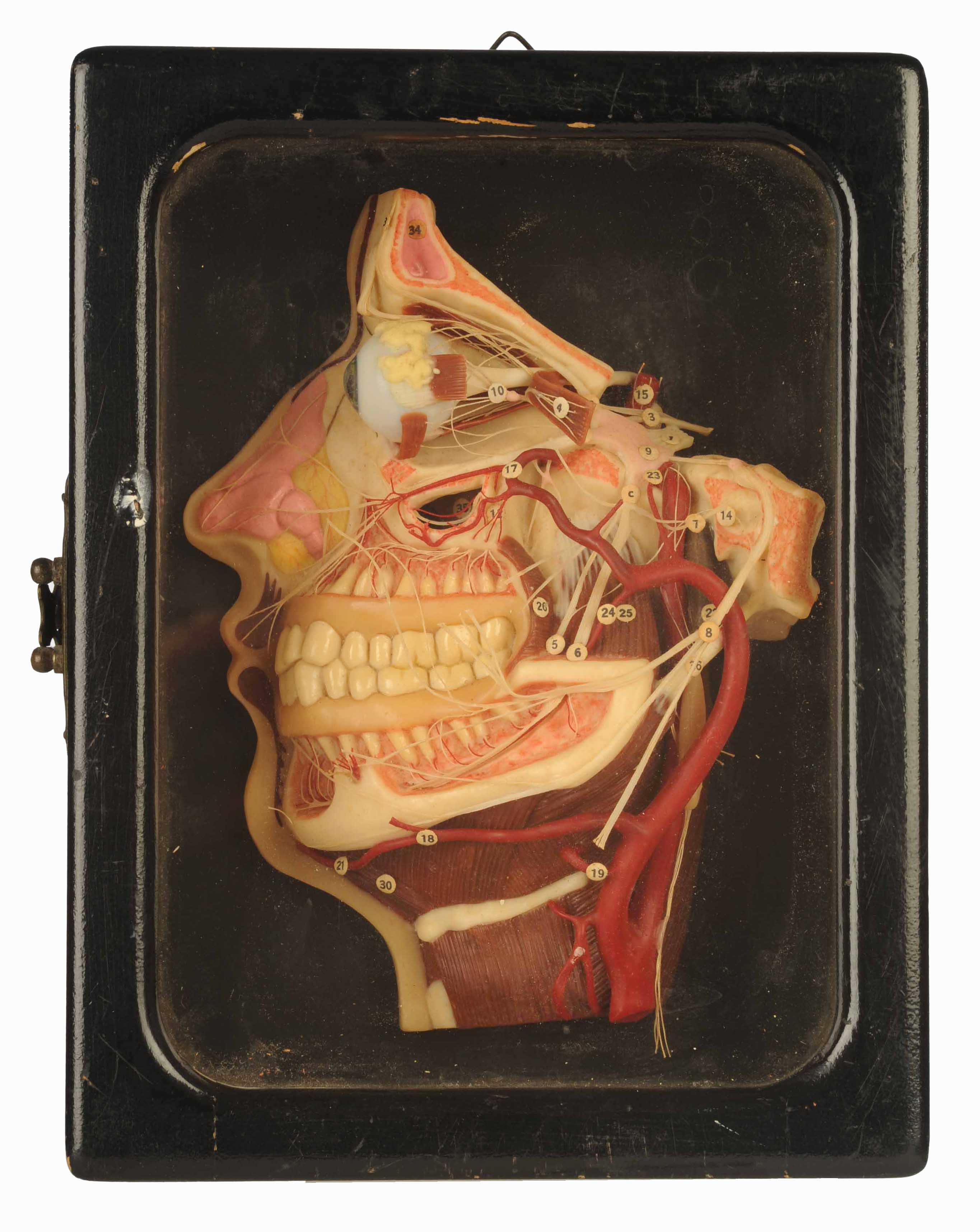
Strangely, this wax model has no legend to explain what each labelled number is indicating. A few of the features that have been identified include:
5: The lingual nerve, which provides feeling in the mouth and the front of the tongue, as well as sending taste signals between the tongue and brain
8: The facial nerve, which controls the facial muscles responsible for smiling, frowning, nose wrinkling and eyebrow-raising
9: The trigeminal ganglion – a big name for the biggest collection of neurons in the body!
25: The masseter muscle, which moves the jaw when chewing
The model was donated by the family of John Thomson Murray, a retired dentist. John’s father had also been a dentist, and the model was originally a feature in his dental office in Cardonald, before he retired in 1968 and passed it on to his son. His son qualified as a dentist the following year, and practiced in Irvine until 2004.
John used the model in demonstrations to his patients, likely to help them understand the cause of their pain, or to explain the treatment they were about to receive. People are commonly afraid of the dentist, or at the very least nervous about having their mouths poked and prodded in. Helping patients to visualise their anatomy could serve as a welcome distraction, and might have been reassuring. Similar models are recorded as having been used in a 1911 Hygiene Exhibition in Dresden, and would have helped to spread medical knowledge among the general population.
Rather than strictly anatomical models, it is more common for dentists to have models that specifically depict oral health concerns. The Lehrmittelwerke Berlinische Verlagsanstalt is known to have sold such models, including one called ‘Diseases of the Teeth,’ but relatively few of their wax models have survived in the century following their production. The model depicts veins, arteries, nerves, and muscles, as well as a few tendons. It is a somewhat curious choice for a dental office, considering so much of the model is dedicated to the circulatory system of the neck and head, while dentistry is primarily concerned with the facial nervous system.
Medical students of all varieties employ different types of rhymes and poems as mnemonic devices to help them remember the complex terminology they study. The entertaining mnemonic the dentist John Thomson Murray used in association with the model, “‘The lingual nerve, it took a swerve around the hyoglossus. “Well I’ll be f****d” said Wharton’s duct. “The b****r’s double crossed us!’” is an example of why this model wouldn’t have been particularly well-suited to his purposes. While the lingual nerve is labelled and present on the model (see number 5), the hyoglossus is shown (the muscle directly below and beneath the jaw bone) but not labelled, and Wharton’s Duct (a tube that brings saliva to the mouth, and is highly relevant in dentistry) is nowhere to be seen. Nevertheless, the model is sure to have been an informative visual teaching aid.
Bibliography
Werner, Nina. Dental moulages and anatomical wax models from the collections of the Dental Institute of the University of Berlin. [Zahnärztliche Moulagen und anatomische Wachsmodelle aus den Sammlungen des Zahnärztlichen Instituts der Berliner Universität (1884-1945).] PhD Diss. 2015.
About the Author

Sara Spitz is a Digital Collections Volunteer with the Curatorial Team. She recently graduated with her MSc in Museum Studies from the University of Glasgow. Her research interests are very broad, but generally tend to focus on medical history. She moved to Scotland from Canada, bringing her two cats with her.”